By: Doug Thal, DVM Dipl., ABVP
Once diagnosed with Navicular Syndrome, immediately pursue treatment and pain management for your horse. However, there is no silver bullet for many horses – no one-time treatment to “cure” the condition. Rather, manage your horse over time using a mix of individualized medical treatments, farriery, plus work and exercise recommendations. Following are a few key general points of treatment:
- Quicker diagnosis and treatment result in higher rates of success.
- Horses with conformational defects are at a disadvantage – treating heavy horses with tiny feet often fails.
- An important part of treatment is that riders/owners/trainers adjust expectations to accommodate the horse’s reduced ability to work, understanding and consider extensive management.
Rest:
We always recommend variable length breaks from intense work like galloping or jumping. This does not mean strict stall rest, as many horses with Navicular Syndrome seem to do better with light to moderate daily exercise.
Farriery: A Cornerstone Of Treatment:
Proper trimming and shoeing on a consistent schedule is a key component of managing this condition. Treatment for a horse diagnosed with Navicular Syndrome must start with a “correct trim”. Shoeing then allows additional mechanical manipulation that provides further help. Radiographs provide vital information to guide the shoeing.
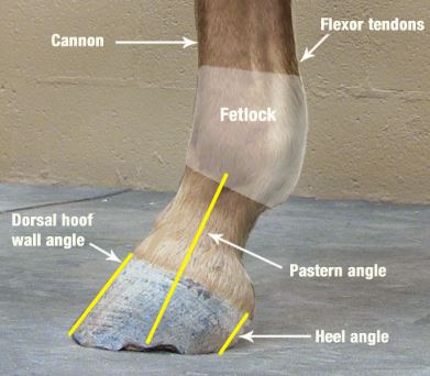
The mechanics of the hoof that are important to consider include:
- Movement of the center of articulation over the center of the hoof,
- Alignment of the hoof-pastern axis,
- Extension of the heel to the base of the frog, if possible, and shortening the breakover.
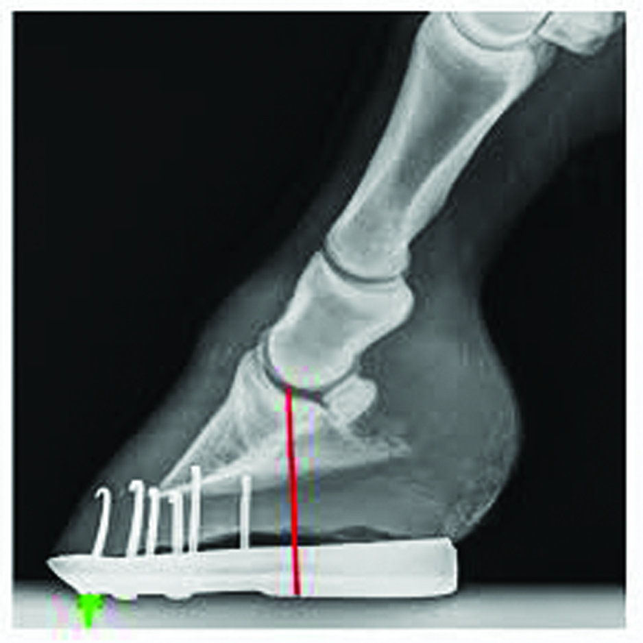
A side (lateral) view of the lower limb is very important to assess hoof/pastern angle and shoeing. This view shows placement of the shoe further forward than might be ideal.
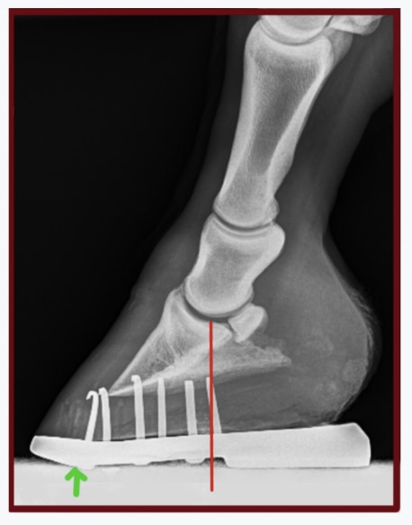
The position of this similar shoe, with more wedge and improved center of rotation has been moved back. Red line indicates center of rotation of the coffin joint, green arrow indicates breakover.
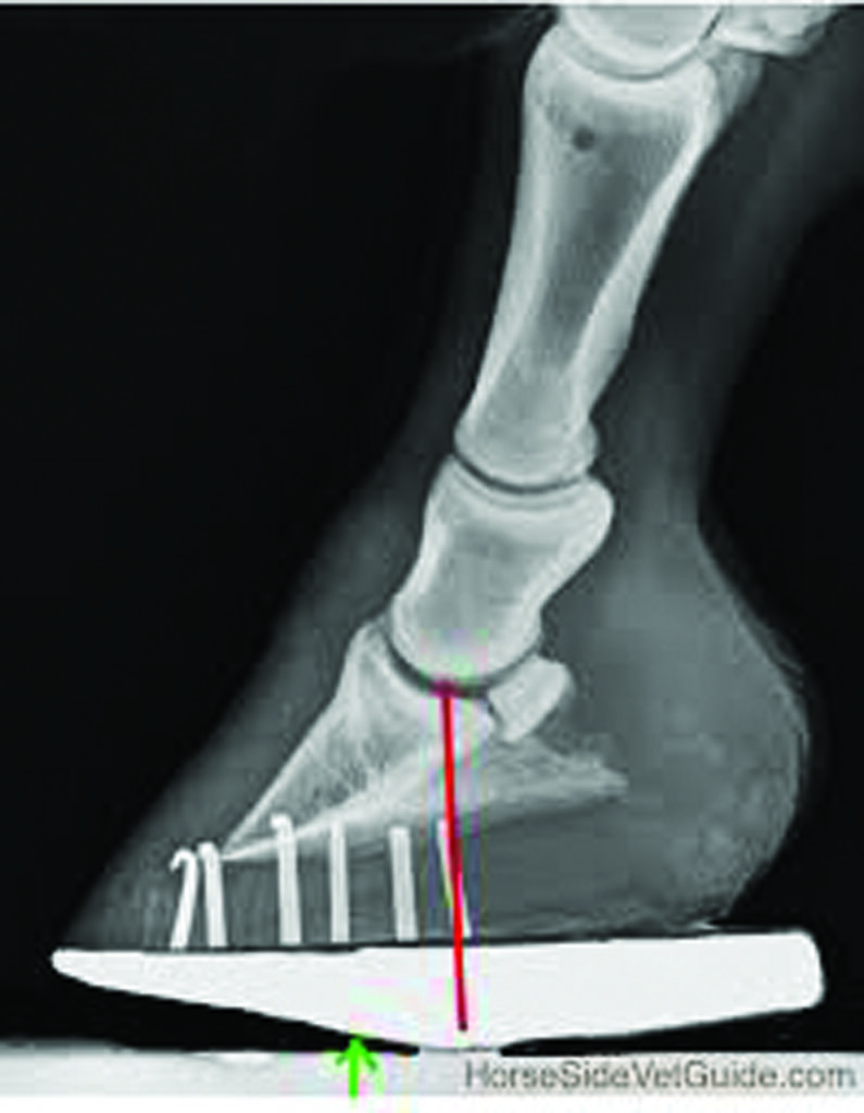
Radiograph showing mechanics of the roller motion shoe on the hoof, identifying one way to address Navicular Syndrome. The red line identifies the center of rotation of the coffin joint. The goal is to relieve stress on the heel and navicular apparatus and thus relieve pain, plus protect the area from direct trauma.
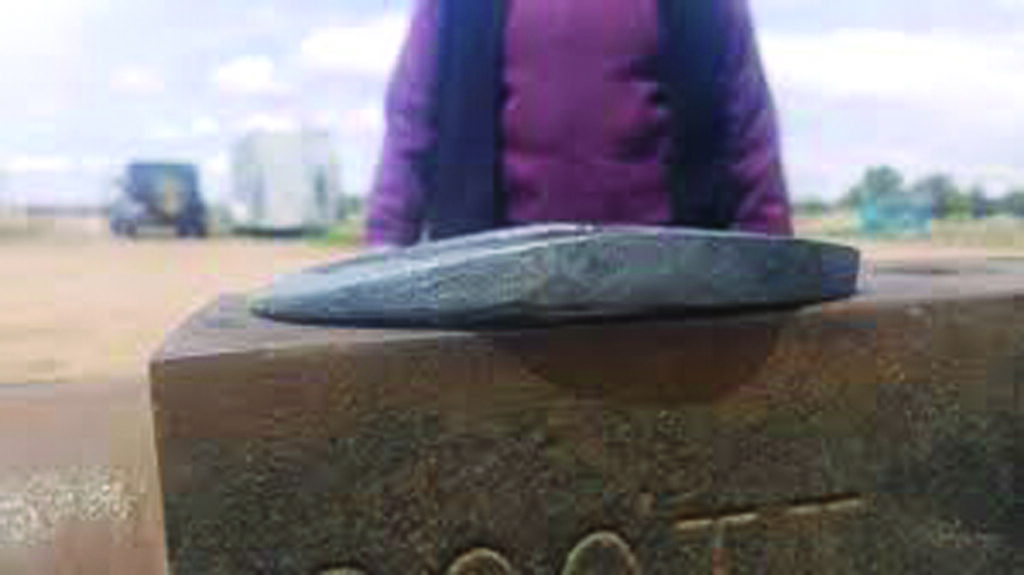
Steel bar-stock was used to construct this roller motion shoe. Though shown upside down, the mechanics can be understood. Toe is to the left.
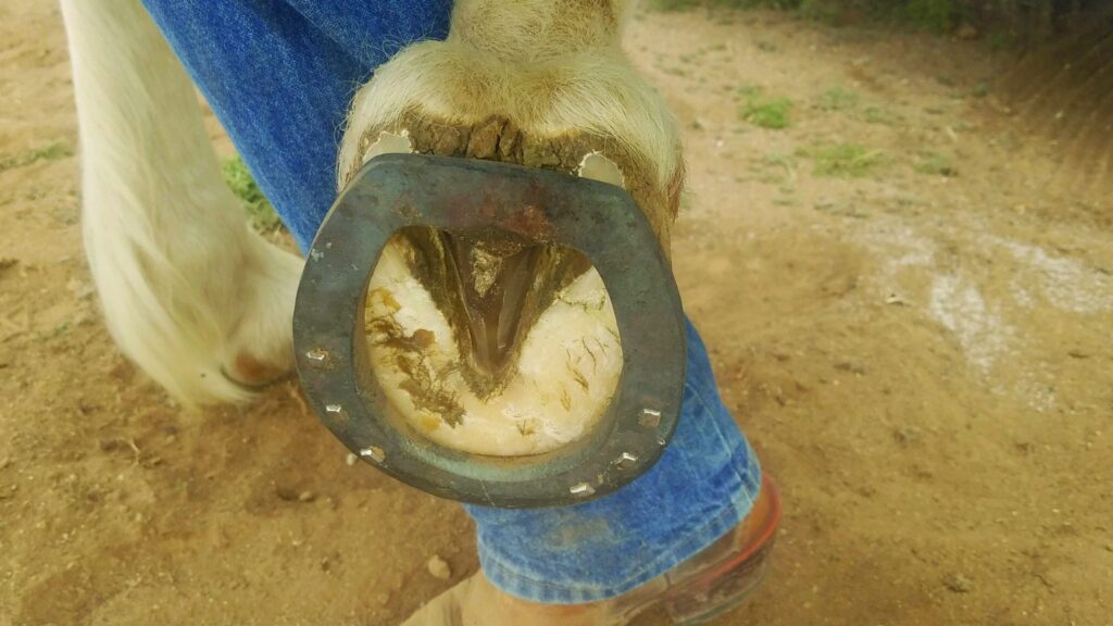
The roller motion bar shoe on the sole of the hoof.
There are many different shoe designs, with the main intent being to wedge the horse to the normal axis, and in soft footing, to reduce over extension of the coffin joint, as this adds load to the navicular. A bar shoe is commonly used to “protect” the heel by decreasing the heel’s sinking into the ground.
Professional disagreements are common about how to manage these horses’ feet. Some suggest removing shoes altogether and managing barefoot. Different farriers will attempt to achieve the mechanical objectives using different materials and techniques.
What is important is following basic principles – and most importantly – that regardless of your plan, the horse is more comfortable and better able to do their job. Working with an experienced veterinarian and experienced farrier gives the horse the best chance of soundness.
Veterinary Treatment:
In the past, all we really knew about this condition was that there was degeneration of the navicular bone visible through x-ray changes in affected horses. The idea was that if this degeneration would stop, then pain coming from the area would stop and the horse would be more comfortable. In many horse, there is some truth to this.
Many horse owners inject medication (usually steroids) into the coffin joint and / or navicular bursa in order to break the cycle of pain and inflammation in the foot. This sometimes provides an improvement in lameness grade. Often it can be difficult to tell just how much this helps, as we often couple injection with changes in shoeing. Repeated injections sometimes show significant and longer lasting improvement. In other cases though, improvement is only transient, suggesting consideration of other strategies.
Systemic medications (oral or injectable medications) are often part of a treatment plan. Historically, treatment of this syndrome employed a wide assortment of medications. When I started practice in the early 1990s, the most commonly used drug was isoxsuprine, a vessel-opening drug (vasodilator) that supposedly encouraged increased blood supply to the bone. Although some horses do improve while on this drug, it was found that in fact it did not increase blood supply to the foot. We really don’t know for sure why isoxsuprine helps in some cases.
These days, a commonly used drug class is the bisphosphonate class (Osphos and Tildren). Bone is a very dynamic tissue, constantly being created and resorbed (taken up). Bisphosphonates works by stopping bone resorption. It makes sense that these might be useful, given that a visible part of Navicular Syndrome appears to be breakdown of the navicular bone itself. As we know though, the disease process is often more complex than that. Some horses with heel pain do respond to these drugs, but some do not. Administer these drugs only under vet supervision, and per the label dosage and interval. If results are poor, other approaches will need to be used instead.
Phenylbutazone (bute), firocoxib, and other Nonsteroidal Anti-inflammatory Drugs (NSAIDS) control pain and inflammation, plus decrease lameness, but because of side-effects are often not a good answer for long-term maintenance. Firocoxib is a safer, though weaker alternative to bute for long-term blunting of pain and inflammation in these horses. Typically, as the disease progresses, NSAIDS present a decreasing solution.
Although widely used, PSGAG (Adequan) and Sodium hyaluronate (Legend) and oral joint supplements are not thought to provide much benefit for horses with Navicular Syndrome.
Extracorporeal Shockwave can be helpful in some cases, particularly those that involve the rear part of the bone (flexor cortex). I have had some very positive results using this therapy in a handful of cases that did not respond to other treatments.
A variety of surgical procedures (like cutting of the navicular suspensory ligaments) can change the mechanics of the navicular and can be helpful in some cases, but are case specific and are not reliable.
For cases that do not respond to treatment, an MRI might be necessary to further define the extent of the injury. Review the navicular bone, the supporting ligaments, the bursa, the coffin joint and / or the deep digital flexor tendon. The information in an MRI image typically provides precise, targeted treatment of these individual structures. Perhaps a tiny tear in the deep flexor tendon could be “cleaned up” arthroscopically, or stem cells injected into it. Long-term rest might also be vital in healing an injury, whereas it wouldn’t be expected to help the horse with more typical bone degeneration. Similarly, the horse with the tendon tear would be unlikely to have a positive response to a bisphosphonate drug.
For totally uncomfortable horses with advanced disease, Palmar Digital Neurectomy (nerving) can be helpful as a last alternative. This procedure involves removal of a segment of each of the heel (palmar digital) nerves in the back of the pastern. Though this provides long-term relief of pain through numbing of the area, it obviously does not correct the condition causing pain. The cycle of destruction in the hoof usually goes on, and in some cases may accelerate owing to more loading of the limb.
Neurectomy has important potential side effects to consider. In cases with a damaged deep digital flexor tendon, ongoing degeneration of that structure through increased wear may eventually cause rupture, requiring euthanasia. Nerve regrowth is common after a few years, with return of pain and lameness. A horse with no sensation in the sole of the hoof may step on a nail and not show lameness, allowing the damage to worsen. These and other disadvantages aside, Neurectomy can be a helpful salvage procedure for some horses, prolonging a quality life in a situation that would otherwise require euthanasia.
Your Role:
As an equine caretaker, your role is just as vital as an experienced vet or farrier’s role.
Be on the lookout for this condition, especially in the predisposed breeds, and especially when you are considering buying a horse.
- Know the basic mechanics and anatomy of the horse’s foot. Know what it should look like and recognize major deviations in a horse’s hoof capsule.
- Get your vet involved early if you suspect lameness.
- Once diagnosed, you will need to administer the treatment and management to the horse as prescribed.
- You will also need to objectively monitor the horse’s response and communicate your observations to your vet.
Over weeks to months, the goal is to see the horse with this condition improve somewhat, with whatever treatment approach is selected. If expected improvement fails to appear, discuss other treatment options with your vet. Depending on the success of initial treatment, try different approaches until you see improvement and the horse is able to do its job adequately and comfortably. In some cases, there is never sufficient improvement, and the horse will not be able to perform its intended use.
Conclusion:
Navicular Syndrome is a very common but complex condition. A complete diagnosis requires a thorough diagnostic approach. Your vet should be very familiar with the many faces of this syndrome, be confident with diagnosis, and know the treatment options. If they are not, they should be willing to refer you to someone who is. The vet MUST work with an experienced farrier to implement the necessary farriery changes. It is the team approach that results in success in this and other conditions of the foot. Whether or not an MRI can be a part of diagnosis depends on the particular case and your resources.
I hope this article is of value to you.
References:
Baxter G. Manual of Equine Lameness: Wiley Blackwell 2006 pp. 225-229
Floyd A. Mansmann R. Equine Podiatry. Saunders 2007
O’Grady S. Strategies for shoeing the horse with palmar foot pain: www.equipodiatry.com/article_palmar_pain